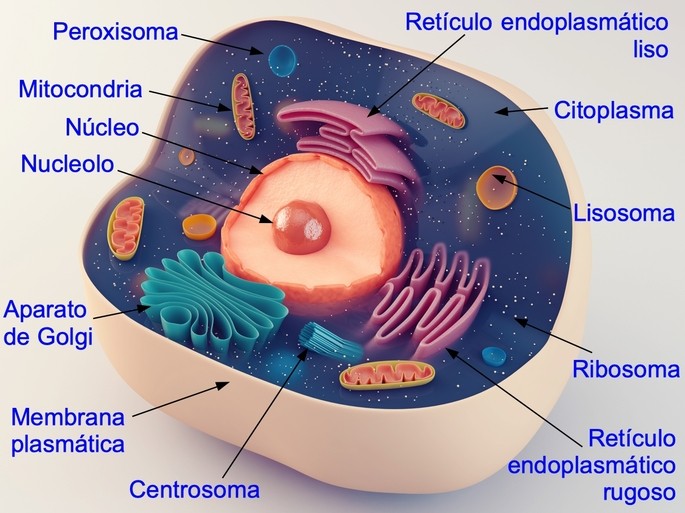
The animal cell is the building block of all animal structures. It is a type of eukaryotic cell, characterized by a nucleus in which the genetic material in the form of deoxyribonucleic acid or DNA is enclosed.
The animal cell presents different parts with specific functions, as shown in the following table:
|
Parts of the animal cell |
Function |
|---|---|
| Plasma membrane |
Protects the interior of the cell |
| Nucleus | Synthesizes DNA and RNA |
| Cytoplasm |
Allows the movement of molecules and organelles. |
| Endoplasmic reticulum |
Assembles and processes proteins |
| Ribosome | Synthesizes proteins |
| Golgi apparatus | Stores and distributes proteins and lipids. Forms vesicles |
| Mitochondria |
Synthesizes ATP (biological energy molecule). Oxidizes fatty acids |
| Lysosome | Digests material ingested by the cell. |
| Peroxisome | Oxidizes fatty acids Synthesizes myelin lipids Removes hydrogen peroxide |
| Centrosome | Organizes and assembles microtubules. |
| Cytoskeleton | Gives structure and support to the cell. Allows cell movement |
Following is a description of each of the parts of the animal cell and what they are used for.
See also: animal and plant cell and plant cell parts.
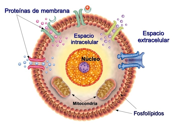
Plasma membrane
The plasma membrane or cell membrane is the outermost part of the cell that limits and closes its contents, separating the extracellular environment from the cell interior. Its structure is fluid and dynamic, consisting of a double layer of lipids, mainly phospholipids and cholesterol, and proteins.
One third of the cell’s proteins are found in the plasma membrane. These are responsible for sensing external conditions or signals and sending that information to the interior so that the cell can respond to the stimulus. Other proteins allow the passage of elements such as sodium and calcium, so that the cell can carry out its activities.
To the plasma membrane is attached the cytoskeleton, in order to maintain the shape of the cell and the movement of intracellular structures.
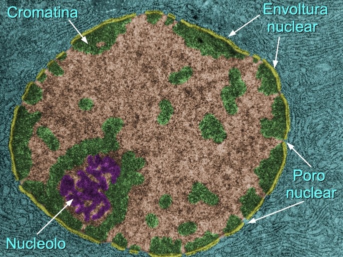
Nucleus
The nucleus is the part of the cell where the genome or genetic information is concentrated as deoxyribonucleic acid (DNA). The functions of DNA and ribonucleic acid (RNA) synthesis, cell division and control of cellular activities are concentrated in the nucleus.
The nucleus can be distinguished by the nuclear envelope, which consists of two membranes with nuclear gaps or pores. During cell division, the nuclear envelope disappears until new cells are formed and rebuilt.
In the nucleus one can also distinguish chromatin, which is nothing more than DNA bound and packaged to nuclear proteins.
Inside the nucleus is the nucleoluspresent in all animal cells, except those that have lost their nucleus, such as red blood cells. The main function of the nucleolus is the production of ribosomes. In growing or cancer cells the nucleolus increases in size.
Cytoplasm
The cytoplasm is the space surrounding the nucleus to the membrane. Within the cytoplasm are immersed the cell’s organelles and microtubule skeleton.
The cytoplasm is composed of:
- The cytosolThe cytosol: the semigelatinous internal fluid in which nutrients and wastes are dissolved.
- Inclusionsare insoluble particles in the cytosol, such as glycogen and fat granules.
- Organellesare “small organs” formed by membranes with specific functions, such as, for example, mitochondria and lysosomes.
- Protein fibers: formed by polymers of small proteins, they include actin microfilaments and tubulin microtubules.
Endoplasmic reticulum
The endoplasmic reticulum is the largest organelle in the cell. It is a constantly changing membrane structure. It participates in the modifications that proteins and lipids undergo during their synthesis and after they are synthesized. It also plays a role in cellular calcium homeostasis.
The endoplasmic reticulum can be divided into:
- The rough endoplasmic reticulum.is a continuation of the nuclear envelope. It consists of stacked sacs of membranes with attached ribosomes, which give it the rough appearance. It is involved in protein synthesis, protein transfer and protein folding.
- Smooth endoplasmic reticulum.:is devoid of ribosomes and is involved in lipid synthesis. Cells such as steroid hormone synthesizing cells and liver cells possess a large amount of smooth endoplasmic reticulum.
Ribosome
Ribosomes are small dense granules of RNA and proteins. Their main function is protein synthesis following the directions of DNA.
There are free ribosomes in the cytoplasm and ribosomes attached to the membrane of other organelles, e.g. the endoplasmic reticulum. Some free ribosomes form clusters of 10 to 20 forming polyribosomes.
Golgi apparatus
The Golgi apparatus or Golgi complex consists of a series of stacked curved sacs that continue the endoplasmic reticulum. It is responsible for receiving proteins synthesized in the rough endoplasmic reticulum, modifying them and packaging them into vesicles for transport to sites where their function is required.
Mitochondria
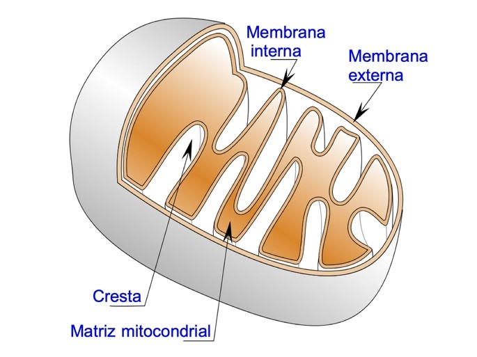
The mitochondrion is a double membrane organelle, the outer mitochondrial membrane and the inner mitochondrial membrane, which delimits the matrix. It is responsible for the production of adenosine triphosphate or ATP, the energy molecule of the cell. In addition, the mitochondrion regulates the cell cycle and apoptosis.
Muscle cells form long networks of mitochondria for rapid and coordinated energy production. In the neuron, mitochondria in the postsynaptic dendrites are larger and interconnected.
Lysosome
Lysosomes are a heterogeneous group of vesicles of different sizes and contents. Their main function is the digestion of the external or internal material of the cell, so they are considered a kind of “cellular stomach”. This is done thanks to several enzymes that degrade proteins, carbohydrates, lipids and nucleic acids.
Lysosomal enzymes are produced in the endoplasmic reticulum, mature in the Golgi apparatus and are transported to the cytoplasm in small vesicles, known as primary lysosomes. Mature lysosomes fuse and divide, making them a dynamic compartment.
Lysosomes exist in all animal cells except the red blood cell. Degradation of endocytosed or autophagocytosed materials takes place within lysosomes that have an acidic pH between 4 and 5. After degradation of the enclosed material, the lysosomes enter a “resting” state.
Peroxisome
Peroxisome is a membranous organelle involved in oxidative metabolism. In mammals, abundant peroxisomes are found in liver and kidney cells.
Peroxisomes are involved in the oxidation of fatty acids, synthesis of myelin lipids, and removal of hydrogen peroxide from cells.
When peroxisomes are malfunctioning or absent, a disease called Zellweger’s syndrome occurs.
Centrosome
Centrosome is a non-membranous organelle that serves as the microtubule organizing center. It facilitates cell motility, polarity, shape maintenance, cell division, vesicle transport. In the interphase or non-dividing phase of the cell, the centrosome is located close to the nucleus.
The centrosome of a mammalian animal cell consists of a protein scaffold surrounding a pair of cylindrical centrioles.
Cytoskeleton
The cytoskeleton is a flexible three-dimensional structure formed by protein filaments. Depending on the thickness of the filament, they are classified into microfilaments (7 nanometers (nm)), intermediate filaments (10 nm) and microtubules (25 nm).
The cytoskeleton maintains the shape of the cell, enables the movement of cilia and flagella, and participates in the intracellular transport of organelles.
You may be interested to see also:
References
Goodman, S. R. (2019). Cell (biology). AccessScience. Retrieved January 25, 2022, from https://doi.org/10.1036/1097-8542.116000
Hettema, E., Gould, S. (2017). Organelle formation from scratch. Nature 542: 174-175. https://doi.org/10.1038/nature21496
Islinger, M., Voelkl, A., Fahimi, D., Schrader, M. (2018). The peroxisome: an update on mysteries 2.0. Histochemistry and Cell Biology 150:443. https://doi.org/10.1007/s00418-018-1722-5
Kurz, T., Terman, A., Gustafsson, B., Brunk, U.T. (2008). Lysosomes in iron metabolism, aging and apoptosis. Histochemistry and Cell Biology 129: 389.